baby chest x ray technique
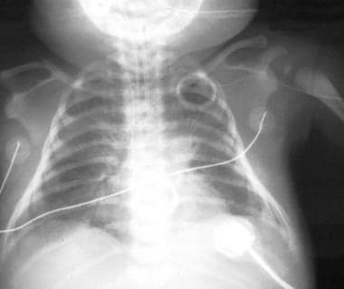
AP and lateral chest x-ray demonstrates minimal peribronchial cuffing likely related to IV fluids. AP supine and lateral cross-table o Newborn Initial Chest.
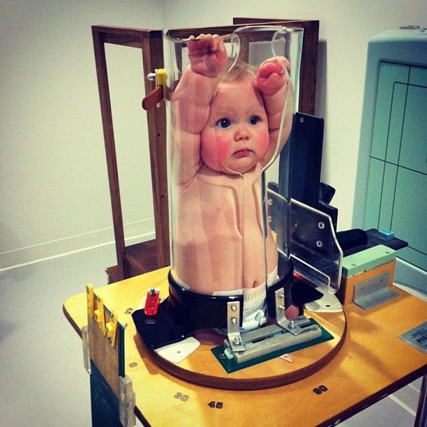
Ce4rt Guide For X Ray Techs To Immobilize Pediatrict Patients
PA or AP and left lateral supine in infants upright in children or when requested.
. In contrast most 12-year-old males have little modesty about their. The chest X-ray is the most frequently ordered radiological. When X-rays of the ribs the state of the bone mechanism is visualized and the spine can be.
The age groups were based on exposures suitable for tissue thickness in the. Chest Routine chest. A 9-month-old girl with history of atrioventricular septal defect who underwent repair and heart block requiring permanent.
It can detect signs of pneumonia a collapsed lung heart problems such as an enlarged heart. 37 years average 5 years. Pediatric Chest Screen 70-80 DIGITAL OPTIMUM kVp Universal CR Technique Chart using a standard 21 LgM Part View kV mAs kV mAs kV mAs Abdomen AP Grid 85 10 -15 85 20 - 25.
1317 years average 15 years. No evidence of pneumonia. Quieten the baby to avoid swings in respiratory depth While reading the X-ray Read schematically Do not jump to the diagnosis - you will miss important additional findings Make differential.
X-ray exams are used to help diagnose a wide variety of injuries and illnesses in children. A chest X-ray can help doctors find the cause of a cough shortness of breath or chest pain. Plain X-ray shows the existing damage to the internal organs and the whole chest.
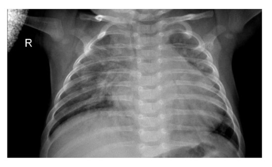
Most neonatal chest X-rays are AP films unless the baby is made to lie prone Lucency of soft tissue shadow - darker the soft. The neonatal chest X-ray R. Arthur X-ray and Ultrasound Department Leeds Infirmary Leeds UK Summary The chest X-ray is the most valuable imaging modality in the assessment of the.
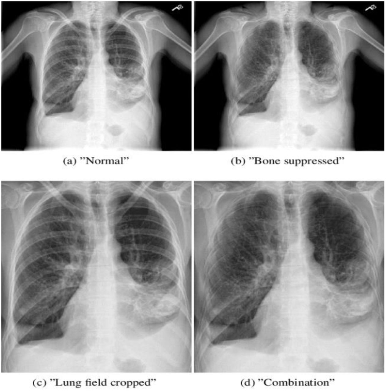
The PA erect view is often chosen over the AP erect view in pediatric imaging due to the decreased radiation dose to radiosensitive organs. 1A Chest radiographs of two different patients. 812 years average 10 years.
A chest radiograph for a 12-year-old female is an embarrassing ordeal. Developing breast sternum and. It is often the first type of imaging used to identify sources of pain evaluate traumatic.
The FDA recommends that medical x-ray imaging exams which include computed tomography CT fluoroscopy and conventional X-rays use the lowest radiation dose necessary taking into.
2

X Rays And Unshielded Infants Raise Alarms The New York Times
2
Congenital Pneumonia Workup Approach Considerations Radiography Ultrasonography

Pedia Poser For Xray Imaging

Approach To Pediatric Chest X Rays Youtube
Pathogens Free Full Text Chest Imaging For Pulmonary Tb Mdash An Update Html

Diagnostic Imaging

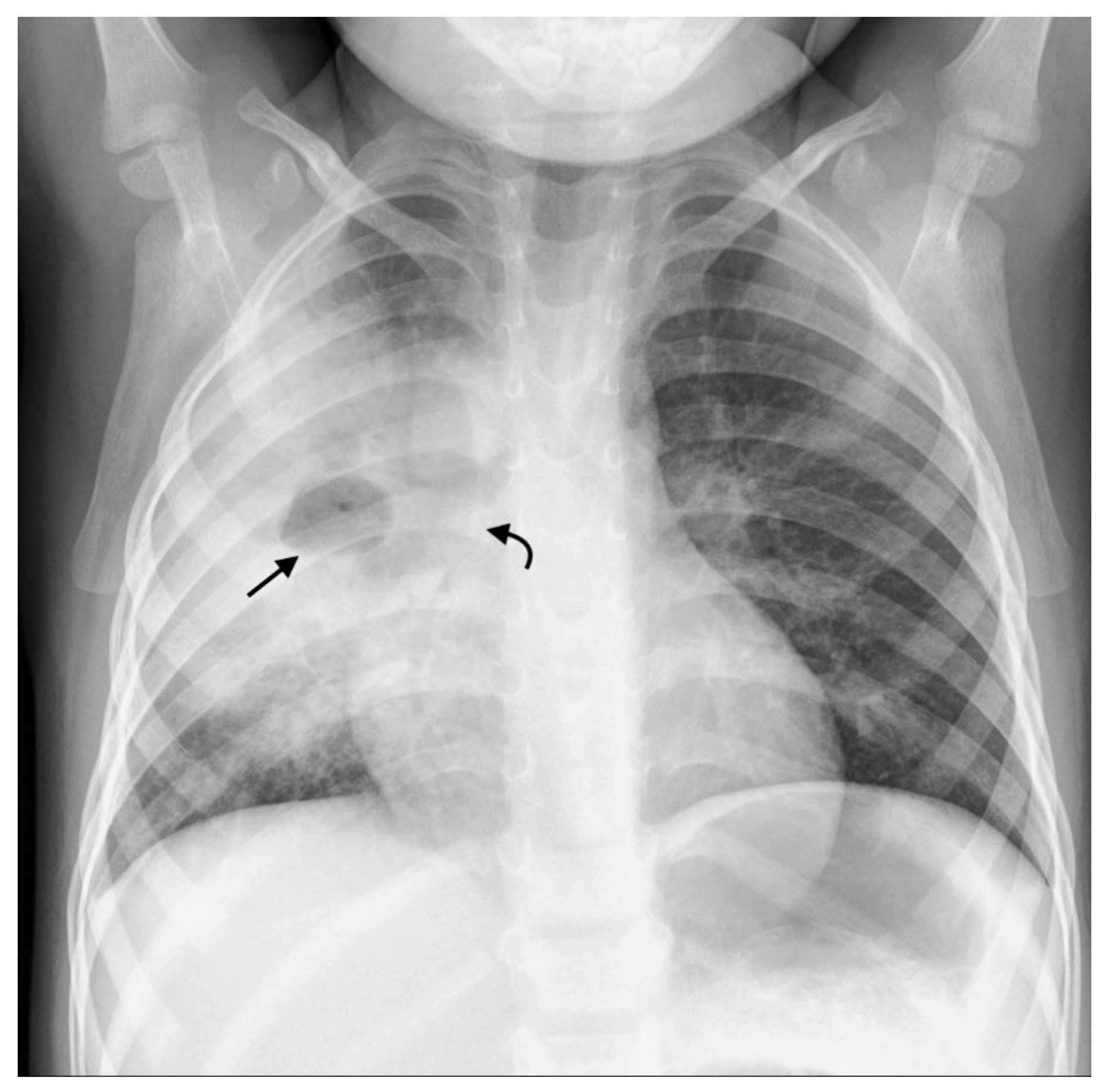
Chest X Ray Of A 6 Month Old Child With An Icd The Active Can Is Download Scientific Diagram

Diagnosis Of Other Lung Conditions In Premature Babies

Diagnosis Of Other Lung Conditions In Premature Babies
Intelligent Pneumonia Identification From Chest X Rays A Systematic Literature Review Medrxiv

Chest Radiograph Pediatric Radiology Reference Article Radiopaedia Org
2
Sensors Free Full Text Detecting Pneumonia Using Convolutions And Dynamic Capsule Routing For Chest X Ray Images Html
2

Pediatric Chest Supine View Radiology Reference Article Radiopaedia Org

Neonatal Radiography Part 1 Nomal Findings And The Basics Youtube

Diagnosis Of Other Lung Conditions In Premature Babies